Brain scans show infants’ brain damage caused by Zika virus

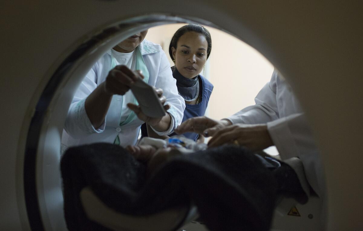
In Campina Grande, Brazil, Amanda dos Santos, 19, watches as medical technicians prepare her baby, 3-month-old Emanuel, before he undergoes a CT scan of his brain.
- Share via
The U.S. Centers for Disease Control and Prevention this week acknowledged that its experts have confirmed public health officials’ worst fears: that Zika virus infection is the direct cause of a massive outbreak of the birth defect microcephaly throughout Brazil.
That grim pronouncement might just be the beginning. The agency said it is exploring whether microcephaly might be the “the tip of the iceberg” in babies born to women who have been infected. For an infected pregnant woman, Zika is increasingly suspected of causing a range of other developmental problems in the child she bears — as well as abnormalities in vision and hearing. And that may be the case even when a newborn’s head is not abnormally small, typically caused by microcephaly.
Even as the CDC drew a definitive line of causation between Zika infection and microcephaly, the British Medical Journal (BMJ) published a detailed study of 23 children (10 girls and 13 boys) born in Brazil’s Pernambuco state to mothers who were infected with Zika during their pregnancies. On 22 CT scans and eight MRI scans, a team of neurologists, radiologists and infant and maternal medicine specialists from across Pernambuco documented severe cerebral damage in virtually all.
FULL COVERAGE: Zika virus outbreak
Above, the CT scan of a baby born with moderate microcephaly shows one of the most curious hallmarks of Zika infection: a near total absence, on the surface of the cortex, of the folds--or convolutions--that massively increase the surface area of a normal human brain (indicated by white arrows). The human gestational brain starts smooth, but begins to develop folds at roughly 23 weeks, as the cortex expands. This condition is called lissencephaly (Latin for “smooth brain”).
In the first image, the black arrow shows another abnormality widely seen in the brains of babies exposed to Zika infection in utero: an extremely underdeveloped corpus callosum, the bundles of fatty tissue that run front-to-back through the middle of the brain, serving as a superhighway for communication between the two hemispheres.
In this CT scan of a baby with severe microcephaly, white arrows point to an extremely stunted corpus callosum and brain stem. The brain stem stretches down to the spinal cord and serves a key function in controlling such involuntary functions as breathing, heart rate, swallowing, blood pressure and consciousness. The cerebellum (short black arrow)--where balance and muscular activity are coordinated--is also abnormally small. Many of the brain’s ventricles in affected babies are abnormally cavernous.
In the image on the right, white specks (black arrows) are calcifications--bony deposits that both crowd out brain cells and disrupt normal connections between them. These damaging lesions were commonly found in babies’ frontal lobes--the seat of attention, planning and higher-order reasoning.
In the top right CT image, frontal lobes that are thickened due to the abnormal migration of neurons--a condition called pachygyria--are seen. Again, the hallmark enlarged ventricles (the long white arrow points to the cisterna magna) and the underdeveloped brain stem (short white arrow) are evident.

A 2-week-old baby girl, born with microcephaly, during a physical therapy session in Campina Grande, Brazil.
The brain abnormalities seen in the BMJ study of Zika-affected babies suggest a “poor prognosis for neurological function,” the authors wrote. Grimly, even the brains of some babies whose head size appeared normal showed stark abnormalities, the authors noted. Imaging revealed these babies’ brain abnormalities were “masked” by enlarged cavities in the spinal-fluid-filled space that normally surrounds the brain’s outer cortex.
Not every pregnant woman infected with Zika has delivered a baby with a brain abnormal in its size, structure and function. But a woman who is infected during pregnancy has an increased risk of having a baby with these health problems, the CDC said.
As a result, the agency urged pregnant women living in or traveling to areas where the Zika virus is being spread by mosquitoes to protect themselves from being bitten and to avoid sexual contact with men who might be infected. And it encouraged women and their partners in such areas to “engage in pregnancy planning and counseling”--possibly delaying pregnancy in light of those risks.
See the most-read stories in Science this hour >>
Follow me on Twitter @LATMelissaHealy and “like” Los Angeles Times Science & Health on Facebook.
MORE ON THE ZIKA VIRUS
Five ways scientists are going after the Zika virus
Even in peak mosquito season, Zika risk is low in California
10 ways to keep yourself safe from Zika, now definitively linked to birth defects


